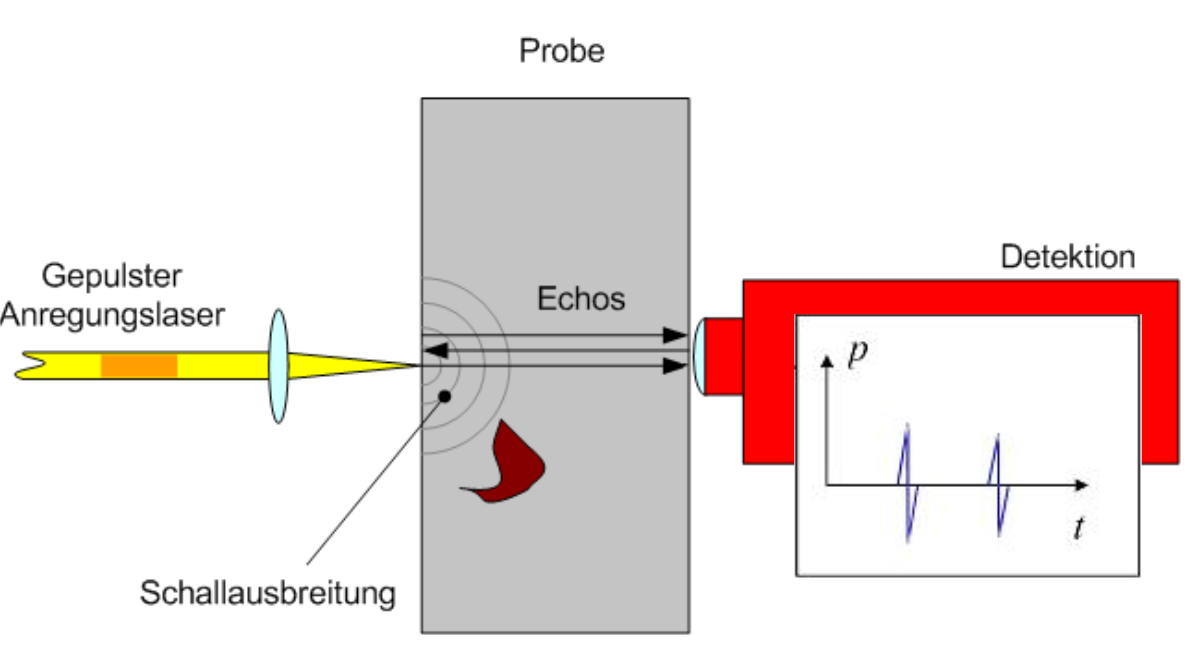
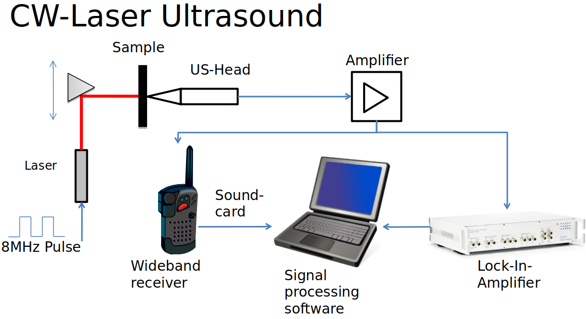
The clinical diagnosis of human dental pulp vitality is the basis for an appropriate therapy of pulp diseases or dental traumata. Most clinical methods currently used to assess pulp vitality are based mainly on the patient's perception. Thermal or electrical stimuli are applied to the respective tooth and pulp vitality is indirectly assessed by the elicited nerve response. Such sensibility responses are subjective and must be communicated to the dentist. In particular with children, older, handicapped, diseased, or patients under anaesthesia this method is not reliable. Numerous other noninvasive technical approaches e.g. those based on Laser Doppler Flowmetry, Photoplethysmography, ultrasound or pulse oximetry have been proposed and are described in literature. However, none of these methods has so far found its way into clinical applications and thus the development of a reliable method for dental pulp vitality testing remains a challenge.
Our approach to the determination of dental pulp vitality utilised the measurement of pulpal blood flow based on the different transmission characteristics of dentine, enamel and blood. Pulp vitality estimation is based on the discrimination between simple light transmission and light modulated by pulsed blood flow. This method was found to be reliable, contactless and does not require tooth-imaging. A major task in this respect was to identify an optimum frequency for which transmission is sufficiently large. Blood absorbs light and its pulsation modulates transmission. Previously only several single frequencies in the visible and infrared (IR) ranges had been applied to the study of dental vitality.
.

