|
DFG-Projekt "Viskograft" |
|
|
|
REM & Histologie 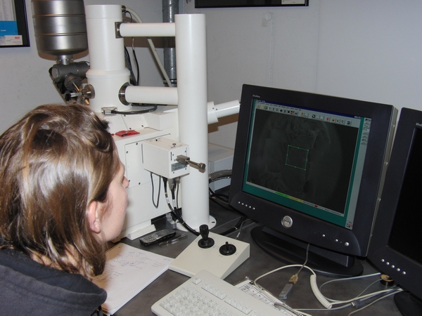 Silvia Mangold am REM der HS.R im Labor von Prof. Dr. Hammer 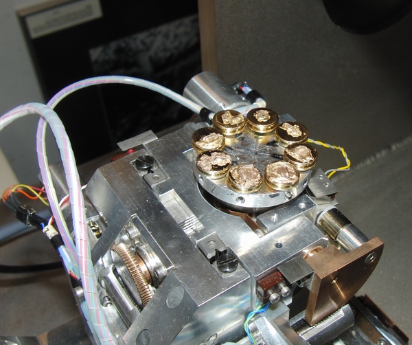 Vergoldete HUV-Proben beim Ladevorgang im REM Abstract Human umbilical
vessels have been recognized as a valuable and widely
available resource
for vascular tissue engineering. Whereas endothelium-denuded human umbilical veins (HUV) have been successfully seeded with a patient-derived neoendothelium, decellularized vessels may have additional advantages due to their lower antigenicity. The present study investigated the effects of three different decellularization procedures on histological, mechanical, and seeding properties of HUV. Vessels were decellularized by detergent treatment (Triton X-100, sodium deoxycholate, IGEPAL-CA630), osmotic lysis (3M NaCl, distilled water), and by peroxyacetic acid treatment. In all cases nuclease treatments were required to remove residual nucleic acids. Decellularization resulted in a partial loss of fibronectin and laminin staining in the subendothelial layer and affected the appearance of elastic fibers. In addition to removing residual nucleic acids, nuclease treatment weakened all stainings and substantially altered surface properties as seen in scanning electron micrographs, indicating additional nonspecific effects. Detergent treatment and osmotic lysis caused failure stresses to decrease significantly. Although conditioned media prepared from decellularized HUV did not severely affect endothelial cell growth, cells seeded on decellularized HUV did not remain viable. This may be attributed to the partial removal of essential extracellular matrix components as well as to changes of surface properties. Therefore, decellularized HUV appear to require additional modifications in order to support successful cell seeding. Replacing the vessels' endothelium only should be considered as an alternative to decellularization when creating tissue engineered blood vessels with non-immunogenic luminal interfaces. Keywords: vascular tissue engineering, small caliber graft, endothelium, biomechanics, human umbilical vein REM 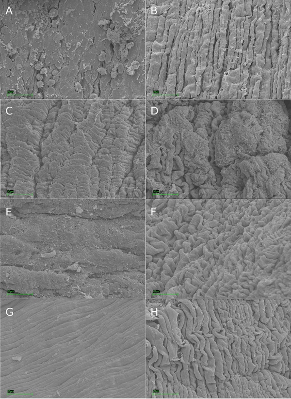 Scanning electron microscopy images of HUV luminal surfaces. (A) native HUV; (B) endothelium-denuded HUV; (C) HUV decellularized by detergents before and (D) after nuclease treatment; (E) HUV decellularized by osmotic lysis before and (F) after nuclease treatment; (G) HUV decellularized by PAA before and (H) after nuclease treatment. Images are representative for 3 independent preparations per condition. Bars indicate 20 μm. Histologie 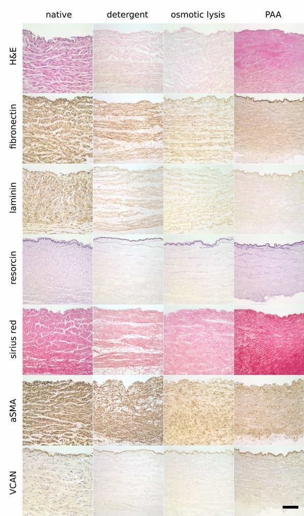 Histological evaluation of HUV decellularization followed by nuclease treatment. See Fig. 1 for explanations. Images are representative for 3-4 independent sample preparations. Bar indicates 100 μm. Veröffentlichung: [15] Mangold, S.; Schrammel, S.; Huber, G.; Niemeyer, M.; Schmid, C.; Stangassinger, M.; Hoenicka, M.: Evaluation of decellularized human umbilican vein (HUV) for vascular tissue engineering - comparison with endothelium-denuded HUV Journal of Tissue Engineering, TERM-12-0055.R1, 2012 |
||
| |
|